

Part 1: Introduction to our brain cells - the neuron
The first part of the introduction to the cells in our nervous system.

I will give an overview here of how neurons are structured, but I will leave the details of how signaling between cells works for a later chapter. Here I will write about what makes this particular cell type special, without going too deeply into cell biology.

The first cell type I will write about is the neuron, and this is probably the cell type that many people think of when talking about "brain cells". In fact, only about half of all brain cells are neurons, the other half are classified as glial cells.1
Introduction to the neuron
Neurons are cells that receive, process, and transmit information in our nervous system. To receive and transmit information in the form of bioelectric and biochemical signals, the cell needs specialized structures for this purpose.
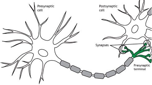
A neuron typically has two different types of projections extending from the cell body, called dendrites and axons respectively. The dendrites are the part of the cell that receives information and branches out like a tree crown with additional small dendritic spines along the dendritic membrane. The axon is a long projection that, unlike the generally shorter dendrites, can extend from millimeters up to about a meter before branching and ending in boutons, also called presynaptic terminals.
In summary, the flow of information goes from the dendrites to the cell body and then through the axon to the presynaptic terminals, and from there to the next cell.

The properties of the axon
Cytoskeleton
The axon itself has, in comparison to the dendrites, more microtubules that are part of the cell's cytoskeleton. In addition to stabilizing the cell, these also function as transport routes for vesicles (containing substances such as neurotransmitters) that are transported along the microtubules through the axon down to the synapses where they can then be released.
In one of the books I'm reading on the subject the transport is compared to how "an insect climbing on a drinking straw", where the insect illustrates a transport protein with a vesicle. In any case, it is hard to forget the description.

Myelin
The speed at which a neuron can transmit signals depends on several properties of the axon, including whether it is myelinated or not. Myelin comes from specific types of glial cells that wrap their cytoplasm around an axon over and over again, which allows signals to travel faster. For example - neurons that transmit information from our muscles to the brain have a thick layer of myelin, while sensory neurons that transmit pain and temperature to the brain are often unmyelinated and conduct signals much slower.
Between the myelinated regions on the axon, the cell's membrane is in contact with the surroundings at the nodes of Ranvier. A recurring analogy is that the signal "jumps" between the nodes and is therefore conducted faster.
Myelin is important for the proper functioning of the nervous system. If the myelin is damaged or breaks down, it can cause disease because signals are not transmitted as quickly or not at all. In the central nervous system, MS (multiple sclerosis) is the most common demyelinating disease.
In short about synapses
A synapse is the area where one neuron (usually the presynaptic terminals) communicates with another neuron (usually the dendritic spines). Near the presynaptic terminals there are zones inside the cell where the vesicles containing neurotransmitters are stored before being released.
The actual synthesis of many neurotransmitters is dependent on enzymes that are transported in vesicles from the cell body along the microtubules as described earlier. Examples of such neurotransmitters can be dopamine, histamine or acetylcholine.
A Brief Overview
So we have gone through the basics of what a neuron looks like and broadly what structures distinguish it from other cells. We have only scratched the surface, and of course there is much more to learn. The intention here is to provide an overview, and of course there are plenty of textbooks that cover the subject in much more detail. I have used the sources listed below.
See you in the next post about glia cells!
Lovisa
Sources:
1. Herculano-Houzel S. (2014). The glia/neuron ratio: how it varies uniformly across brain structures and species and what that means for brain physiology and evolution. Glia, 62(9), 1377–1391. https://doi.org/10.1002/glia.22683
Picture sources:
Picture 1: Brett Szymik. (2011, May 03). Neuron Anatomy. ASU - Ask A Biologist. 28 maj, 2024 https://askabiologist.asu.edu/neuron-anatomy
Picture 2: Wikipedia.
Picture 3: Henley, Casey (2021). Foundations in neuroscience. Chapter 8: Synapse structure. Fig 8.1.https://openbooks.lib.msu.edu/neuroscience/chapter/synapse-structure/
Books I based this text on:
G.A. Mihailoff, D.E. Haines, Chapter 2 - The Cell Biology of Neurons and Glia, Editor(s): Duane E. Haines, Gregory A. Mihailoff, Fundamental Neuroscience for Basic and Clinical Applications (Fifth Edition), Elsevier, 2018, Pages 15-33.e1, ISBN 9780323396325,https://doi.org/10.1016/B978-0-323-39632-5.00002-5.(https://www.sciencedirect.com/science/article/pii/B9780323396325000025)
Luo, L. (2020). Principles of Neurobiology (2nd ed.). Garland Science. https://doi.org/10.1201/9781003053972